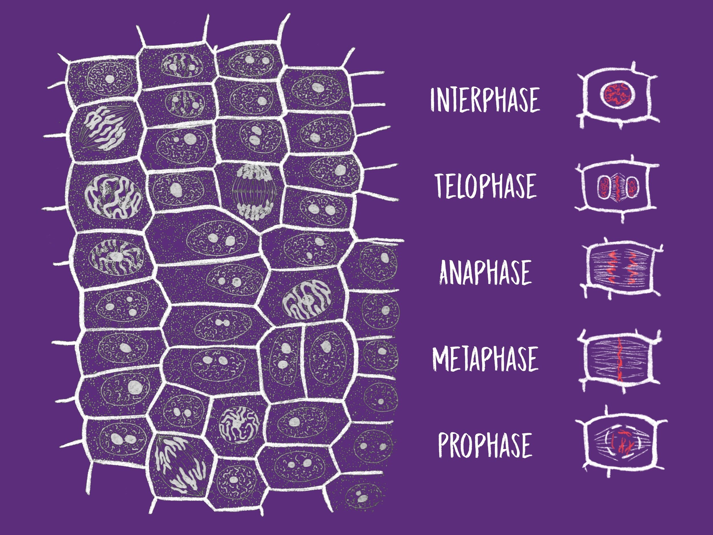
Mitosis in Onion Root Tips — DataClassroom
From Retail to Warehousing, We Have Barcode Labels for All Your Needs! Ensure Accurate Inventory Management with Our Barcode Labels! Shop Now!
Onion Plant Cell Under Microscope Labeled / Onion Cells Onion
Onion Skin Cells - Investigation Objectives: Observe plant cells Materials per student (or team): • onion • microscope • clean microscope slide and cover slip • tweezers • eye dropper • stain - methylene blue staining solution (iodine solution can be used as an alternative stain) • paper towel Procedures: 1.
PPT Amoeba PowerPoint Presentation, free download ID6663278
Label the cell wall, middle lamella, plasmodesmata, and chromoplasts. You are encouraged to identify and label other cell components, such as the nucleus and nucleolus, if they are visible. A potato is a modified part of the plant called a tuber. Much like an onion, a tuber is a part of the plant--this time the stem--adapted for storing starch.
Microscope Onion Cell Labeled Micropedia
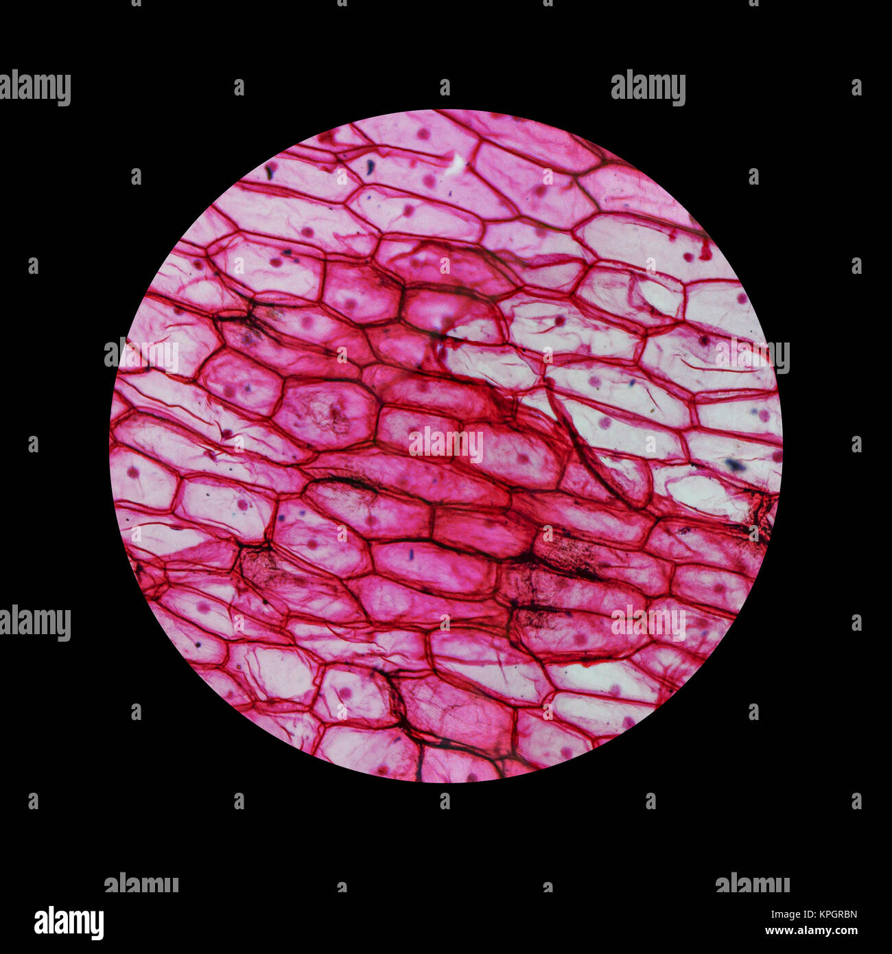
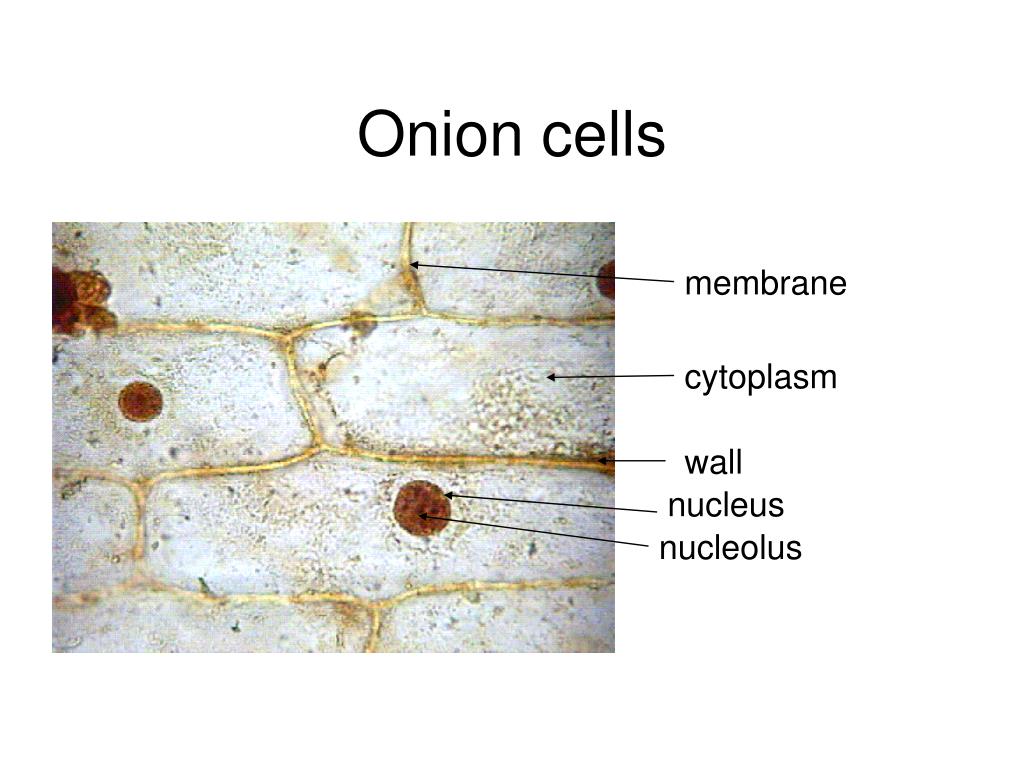
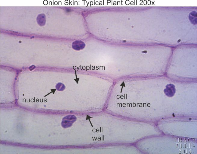
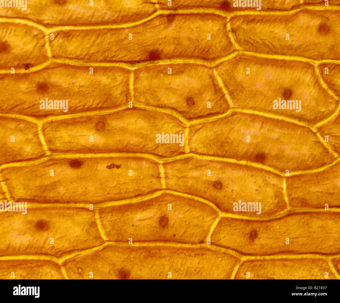
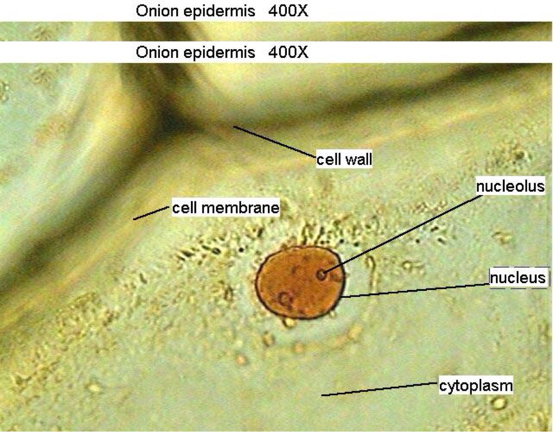
An onion is a multicellular (consisting of many cells) plant organism. As in all plant cells, the cell of an onion peel consists of a cell wall, cell membrane, cytoplasm, nucleus and a large vacuole. The nucleus is present at the periphery of the cytoplasm. The vacuole is prominent and present at the centre of the cell.

Onion Root Tip Mitosis
Updated July 11, 2019 By Peg Robinson Onions have a long history of human use, originating in southwestern Asia but having since been cultivated across the world. Their strong odor — actually a defense mechanism — and unique structure belie a complex internal makeup, composed of cell walls, cytoplasm, and the vacuole.

Onion cells containing onion, cell, and cells HighQuality Nature
To answer your question, onion cells (you usually use epithelial cells for this experiment) are 'normal' cells with all of the 'normal' organelles: nucleus, cytoplasm, cell wall and membrane, mitochondria, ribosomes, rough and smooth endoplasmic reticulum, centrioles, Golgi body and vacuoles.

Labeled Onion Cell In Microscope
The Onion Peel Cell Experiment is a popular and educational activity used to observe and understand the structure of plant cells. This experiment focuses on the onion, a eukaryotic plant known for its multicellular composition. As we delve into this experiment, we explore the essential components that make up a cell, the building blocks of life.

Biology LectureHub
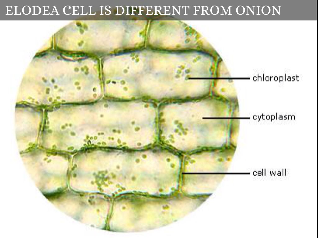
Onion Cells Under a Microscope ** Requirements, Preparation and Observation The bulb of an onion is formed from modified leaves. While photosynthesis takes place in the leaves of an onion containing chloroplast, the little glucose that is produced from this process is converted in to starch (starch granules) and stored in the bulb.
Microscope Onion Cell Labeled Micropedia
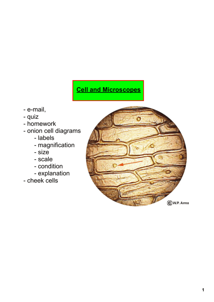
The Onion and Cheek Cell Lab Background: Onion tissue provides excellent cells to study under the microscope. The main cell structures are easy to see when viewed with the microscope at low power. For example, you will observe a large circular nucleus in each cell, which contains the genetic material for the cell.

Onion_Cells
Onion Cell Lab Power __________ Total Magnification __________ After you have completed the rest of this lab come back to this cover page DRAW & LABEL AN ONION CELL WITH ALL THE PARTS / ORGANELLES YOU OBSERVE UNDER 40X. Purpose: To observe and identify major plant cell structures and to relate the structure of the cell to its function.

[DIAGRAM] Labeled Onion Cell Diagram
Start studying Onion Cell Labelling. Learn vocabulary, terms, and more with flashcards, games, and other study tools.
Microscope Onion Cell Labeled Micropedia
All living organisms are made up of cells. Cells are the smallest part of a living organism and are around 0.01 mm - 0.03 mm long. To look at a cell close up a microscope needs to be used.

Onion Cells Size Does Matter
Show your students how to prepare a slide from an onion, view onion cells under the microscope, and observe the structure. Then teach them how to draw and label the structure of an onion cell including the nucleus and cell wall with this great investigation resource. Show more.

Onion Root Tip Mitosis Labeled Diagram
What do onion cells look like under the microscope? Studying cell tissues from an onion peel is a great exercise in using light microscopes and learning about plant cells, since onion cells are highly visible under a microscope, especially when stained correctly.
Onion Plant Diagram
Conclusion Objective The main objective of performing the onion peel cell experiment is to observe the arrangement and structural components of the onion epidermis. The following facts about the onion peel cell experiment play a significant role in educating students:
Onion Plant Cell Under Microscope Labeled / Onion Cells Onion
Overview. Students make slides of cells from an onion skin and an Elodea leaf to observe under a microscope, and learn that all organisms are composed of cells.. This activity is from The Science of Microbes Teacher's Guide, and is most appropriate for use with students in grades 6-8.Lessons from the guide may be used with other grade levels as deemed appropriate.
