
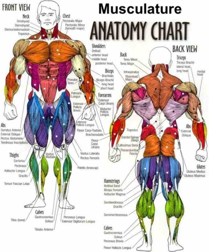
Muscles Chart Anatomy Diagram
Each skeletal muscle is an organ that consists of various integrated tissues. These tissues include the skeletal muscle fibers, blood vessels, nerve fibers, and connective tissue. Each skeletal muscle has three layers of connective tissue (called "mysia") that enclose it and provide structure to the muscle as a whole, and also.

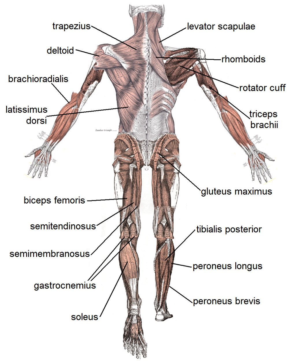
Back Muscles Diagram Labeled Labeled Muscle Diagram — UNTPIKAPPS We
Roll your mouse over any muscle in the diagram below to learn its name. You can click on any highlighted muscle to view a more detailed image of the muscle and a description of what it does.

Muscle Chart The Image Kid Has It!
Each skeletal muscle is an organ that consists of various integrated tissues. These tissues include the skeletal muscle fibers, blood vessels, nerve fibers, and connective tissue.

Pin on Muscular System
THE MUSCULAR SYSTEM COMPILED BY HOWIE BAUM 1 Muscles make up the bulk of the body and account for 1/3 of its weight.!! Blood vessels and nerves run to every muscle, helping control and regulate each muscle's function. The muscular system creates body heat and also moves the: Bones of the Skeletal system Food through Digestive system
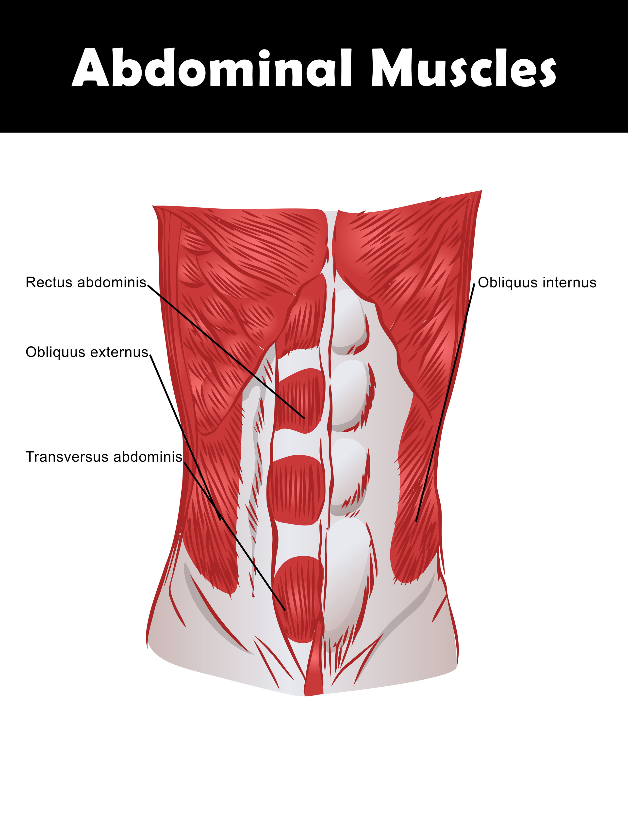
Good Abdominal Exercises
Muscular System Anatomy, Diagram & Function | Healthline human body maps muscular system Muscular The primary job of muscles is to move the bones of the skeleton, but muscles also.

Muscles Labeled Front And Back / 34 Muscle Label Labels Design Ideas 2020
The Female Muscular System Laminated Anatomical Chart. Anatomical Chart Company. Retail Price $23.99. Today's Price $19.99. The muscular system is among our most vital systems. Study it with Anatomy Warehouse's collection of anatomy charts and posters, available at the best prices every day.

muscular system diagram for kids
Muscles and muscle tissue Author: Declan Tempany BSc (Hons) • Reviewer: Christina Loukopoulou MSc. Last reviewed: October 30, 2023 Reading time: 26 minutes Recommended video: Skeletal muscle tissue [12:25] This type of tissue is found in skeletal muscles and is responsible for the voluntary movements of bones. Skeletal muscle
Muscles Of The Torso Model (Anterior And Posterior View) Extending
Muscle is one of the four primary tissue types of the body, and the body contains three types of muscle tissue: skeletal muscle, cardiac muscle, and smooth muscle (Figure 10.2).All three muscle tissues have some properties in common; they all exhibit a quality called excitability as their plasma membranes can change their electrical states (from polarized to depolarized) and send an electrical.
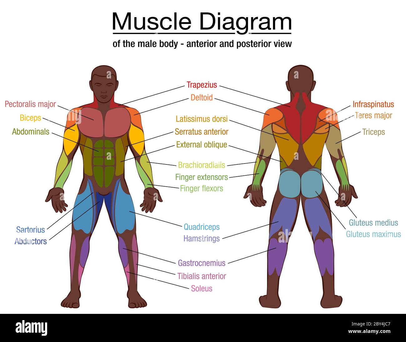
Muscle diagram, most important muscles of an athletic black man
We've created muscle anatomy charts for every muscle containing region of the body: Upper limb Lower limb Head and neck Trunk wall Each chart groups the muscles of that region into its component groups, making your revision a million times easier. For example, upper limb muscles are grouped by shoulder and arm, forearm and hand.
Labeled Body Muscle Diagram
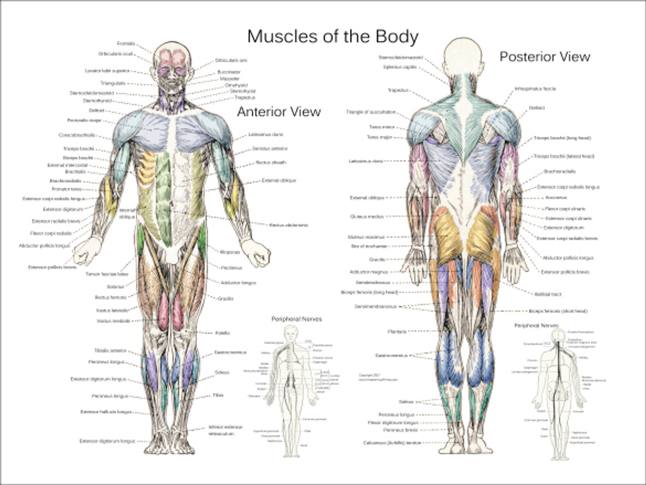
Muscle Charts of the Human Body For your reference value these charts show the major superficial and deep muscles of the human body. Superficial and deep anterior muscles of upper body Superficial and deep posterior muscles of upper body Anterior and posterior muscles of the upper arm Anterior and posterior muscles of the lower arm

Muscle Anatomy Poster Anterior
Osmosis High-Yield Notes. Muscular system anatomy and physiology. Sliding filament model of muscle contraction. Muscle contraction. Neuromuscular junction and motor unit. Osmosis Muscles high-yield notes offers clear overviews with striking illustrations, tables, and diagrams. Make learning more manageable.

25 best images about Muscular Anatomy for Pilates on Pinterest Thighs
Muscles Brought to you by Merck & Co, Inc., Rahway, NJ, USA (known as MSD outside the US and Canada)—dedicated to using leading-edge science to save and improve lives around the world. Learn more about the MSD Manuals and our commitment to Global Medical Knowledge .

Muscles Diagrams Diagram of muscles and anatomy charts
This article is concerned with the skeletal muscles of the human body, with emphasis on muscle movements and the changes that have occurred in human skeletal musculature as a result of the long evolutionary process that involved the assumption of upright posture.

human anatomy muscles labeled
A muscle of the medial thigh that originates on the pubis. It inserts onto the linea aspera of the femur. It adducts, flexes, and rotates the thigh medially. It is controlled by the obturator nerve. It pulls the leg toward the body's midline (i.e. adduction) Biceps brachii An upper arm muscle composed of 2 parts, a long head and a short head.
4 human body muscles labeled Biological Science Picture Directory
Muscle diagrams are a great way to get an overview of all of the muscles within a body region. Studying these is an ideal first step before moving onto the more advanced practices of muscle labeling and quizzes. If you're looking for a speedy way to learn muscle anatomy, look no further than our anatomy crash courses .

Muscle Diagram Most Important Muscles Of An Athletic Male Body Anterior
Muscular System Anatomy and Physiology Updated on October 19, 2023 By Marianne Belleza, R.N. Dive into the ultimate study guide for the muscular system, where anatomy and physiology converge. Nursing students, elevate your understanding and master the art of human motion with every page turn. Table of Contents Functions of the Muscular System
